Scientists take huge step forward in mapping all 37 trillion cells in the human body
The human body contains around 36 trillion to 37 trillion cells, and researchers are mapping out where every one of those cells lives.
Scientists with the Human Cell Atlas (HCA), an international research consortium, have profiled 100 million cells from more than 10,000 people around the world. Working in over 100 countries, the researchers aim to pinpoint similarities and differences in the cells of people from different demographics and genetic backgrounds.
By 2026, the team plans to unveil an atlas of the whole human body that details the location, identity and function of each cell at different stages of life. That atlas will just be a first draft; later iterations could include data from billions of human cells, the scientists project.
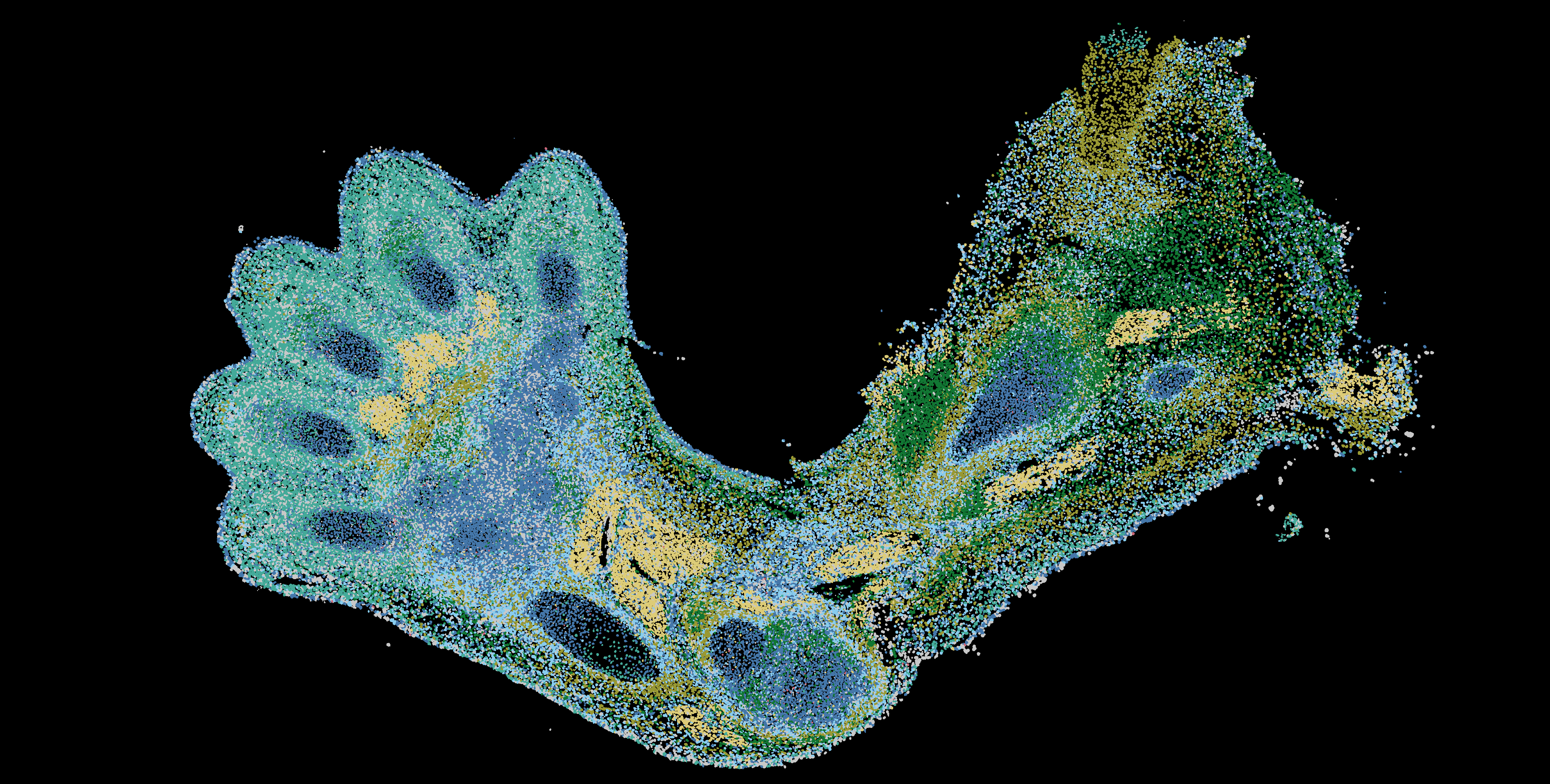
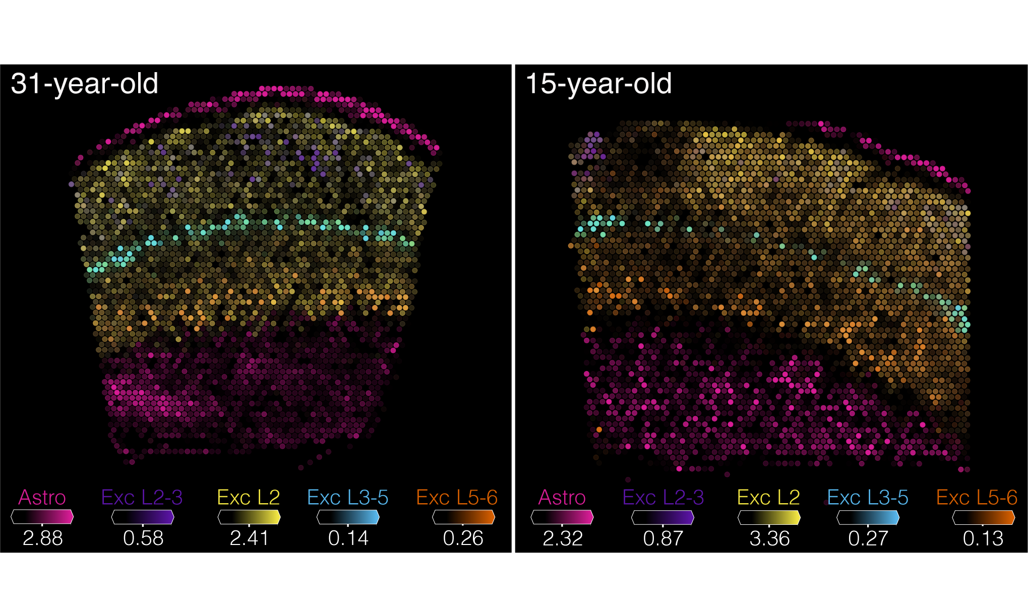
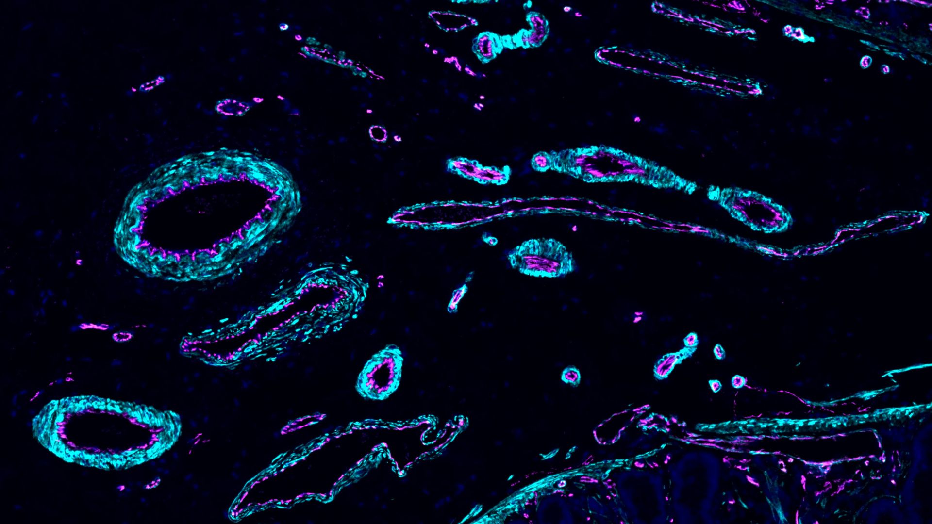
Now, HCA scientists have just dropped more than 40 papers that will aid in the construction of the groundbreaking first draft of their atlas. The research trove, published Wednesday (Nov. 20) in several Nature journals, charts cells in many organs and organ systems — including the lungs, brain and skin — and describes the advanced computational tools needed to crunch all those data.
Related: Most detailed human brain map ever contains 3,300 cell types
Aviv Regev, a founding co-chair of the HCA, compared the advance to leaps in traditional cartography. Imagine going from having only 15th-century maps of the world to having Google Maps, complete with detailed topographies, street views and dynamic traffic patterns.
“So that is the leap that we have done — moving from maps that look as crude as that to maps that are the resolution of the Google Map,” Regev said at a news conference Tuesday (Nov. 19). “But we still have work to do.”
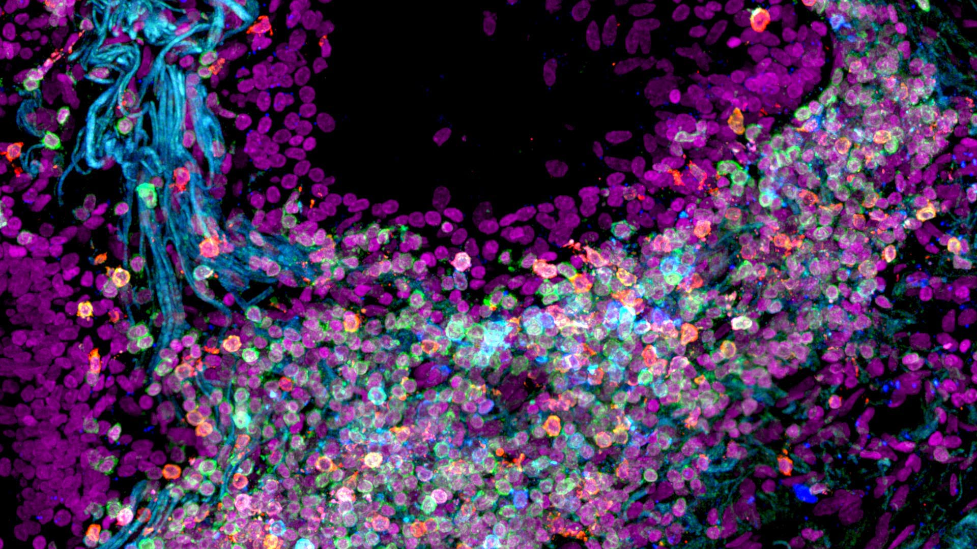
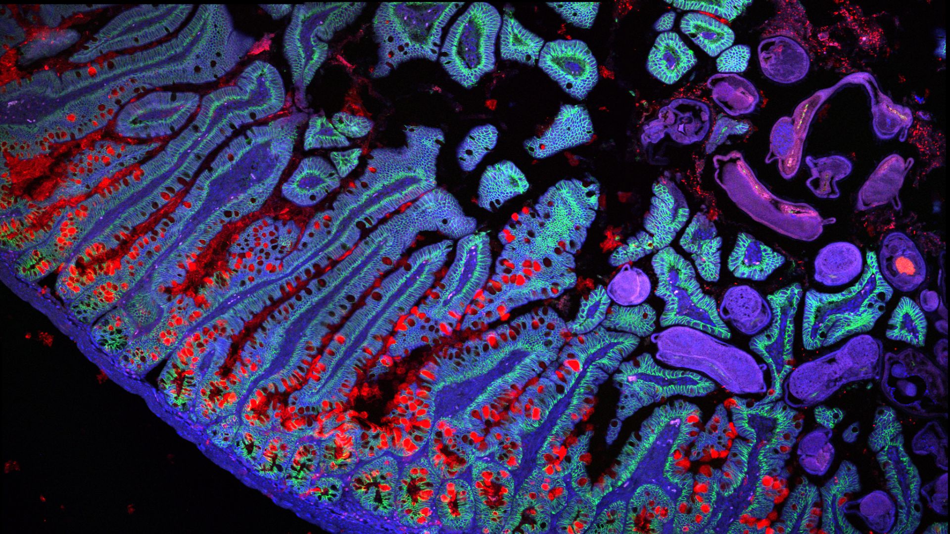
The new research includes a detailed map of the digestive tract, running from the esophagus to the colon. The researchers mapped a healthy digestive tract based on 1.1 million cells sampled from nearly 190 people. They also compiled data from people with different gastrointestinal conditions, including ulcerative colitis and Crohn’s disease. Through this work, they uncovered a type of cell that seems to contribute to the inflammation in these diseases, likely by summoning immune cells.
“Intestinal inflammation can cause cells to undergo metaplasia, a shift from one cell type to another,” Itai Yanai, scientific director of the Applied Bioinformatics Laboratories at NYU Langone Health, wrote in a commentary. With data from both healthy and diseased guts, the researchers were able to pinpoint which stem cells gave rise to the “metaplastic” cells, Yanai said. After transforming, the metaplastic cells then spurred more inflammation, the research suggested.
In other papers, researchers opened a window into early human development, revealing how the placenta develops and the skeleton starts to form in the first trimester of pregnancy. The latter study revealed never-before-seen states that cells enter as they prepare to form the skull. The researchers also investigated genes that might be involved in craniosynostosis, a birth defect in which the soft spots of the skull fuse too soon.
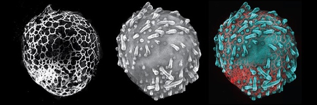
Other papers focused on “organoids,” miniature versions of human organs grown in the lab. One looked at brain organoids, which mimic developing brains. Different labs use various strategies to grow organoids, but that raises questions about which method produces the best model — which brain organoid best mimics an actual brain? The scientists compared maps of human brains to those of organoids, finding that, at least up to the second trimester, the organoids match fetal brains fairly closely. Open questions remain about the third trimester.
Related: Scientists launch amazing ‘atlas’ of embryos, showing how cells move and develop through time
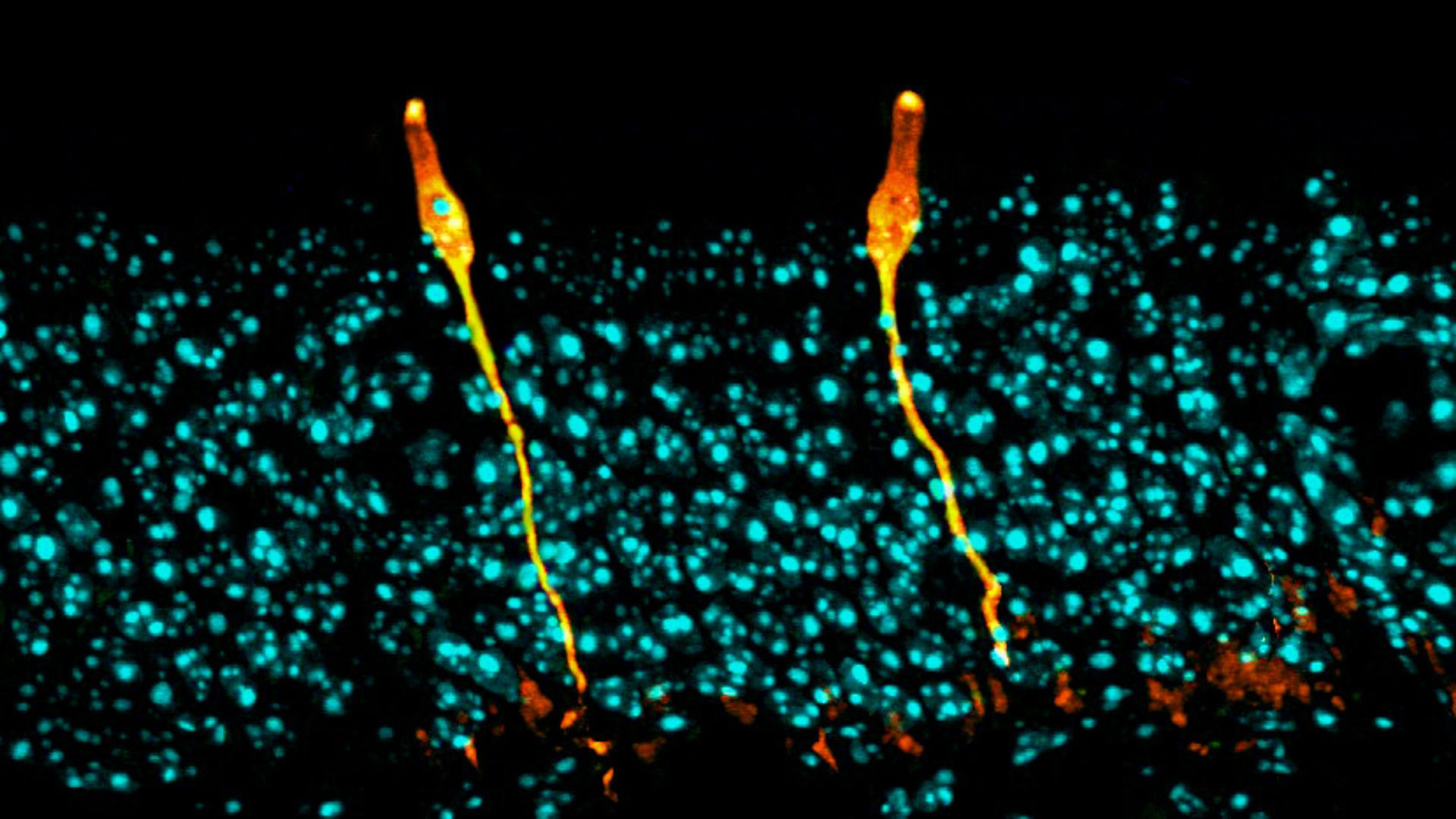
Another lab ran a similar study looking at skin organoids, to see how closely they resembled real skin.
The atlas helps researchers come up with “better recipes” for organoids, Muzlifah Haniffa, a member of the HCA organizing committee, said at the news conference.
But “the information kind of flows both ways,” Sarah Teichmann, an HCA co-chair, added, because the organoids also reveal subtle details of what’s happening inside the body. Scientists can “poke the cells, perturb the cells” in ways that wouldn’t be possible in human subjects, she said. Thus, making true-to-life organoids can help reveal how diseases arise and which drugs might effectively treat them.
Taken together, the more than three dozen HCA papers represent a major step forward. Meanwhile, data previously published by the consortium have already fueled new discoveries: Work published in 2020 helped reveal unexpected tissues that were vulnerable to COVID-19, and a map of the human lung revealed a new type of cell — the ionocyte — that may play a key role in cystic fibrosis.
“Collectively, the atlases have the potential to constitute a resource that others might be inspired to explore and compare with other biological contexts, such as different species and rare diseases,” Yanai wrote. “Researchers might then discover aspects of the human body that cannot yet be imagined.”
Ever wonder why some people build muscle more easily than others or why freckles come out in the sun? Send us your questions about how the human body works to community@livescience.com with the subject line “Health Desk Q,” and you may see your question answered on the website!
Source link

